


Martin Lardy


Vangl2 short and long
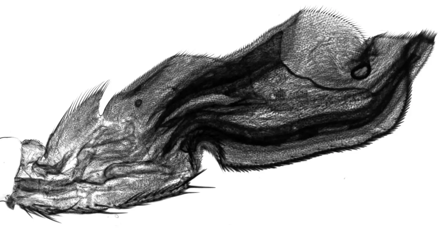
Translation starts with a Methionine: True, but not always, as revealed in a study of the PCP component Vangl2.
The brain: a permanent work in progress
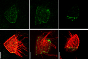
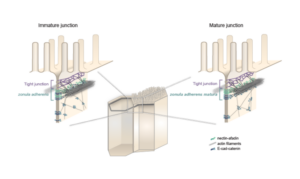
Lenne and Le Bivic teams show that intestinal adherens junctions are very different from the textbook picture.
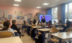
IBDM Marseille inspires young minds: engaging primary school children on childhood cancer (“Contre le cancer, j’apporte ma pierre”) and interacting with high school students through immersive experiences (DECLICS).
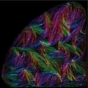
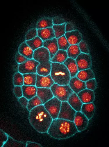
Epithelial tissues under tension: a study explores how individual cells deform and respond to forces.
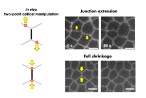
Lenne group, together with 3 other groups, Merkel (CNRS), Trivedi (EMBL), and Ruprecht (CRG) – embark on BREAKDANCE
Organizing the organizers
A new perspective to tackle the very complex cell polarity field.
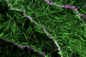
Self-organisation of human muscles in a dish
Human muscle cells self-organise into defined fiber bundles in vitro even without the presence of external cues !
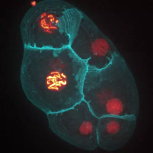
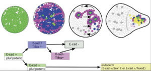
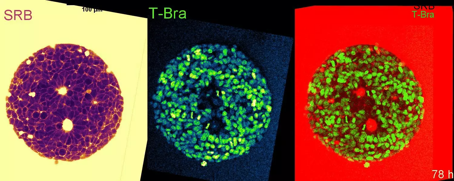
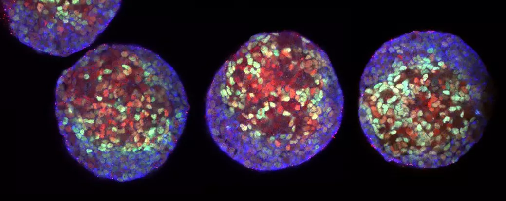
Cell-state transitions and collective cell movement generate an endoderm-like region in gastruloids
The Lenne team published in Elife: using gastruloids –3D aggregates of mouse embryonic stem cells- they study at cellular resolution the specification of the endoderm.
7 IBDM teams have received grants from ANR
7 IBDM teams have received grants from the Agence Nationale pour la Recherche (ANR) in 2021. Congratulations to Vincent Bertrand, Harold Cremer, Pascale Durbec, André Le Bivic, Pierre-François
Pierre-Francois Lenne elected as EMBO member
Heidelberg, 7 July 2020 – EMBO has bestowed upon 63 leading scientists the lifetime honour of EMBO Membership in recognition of their remarkable achievements in the life sciences, it was announced today.
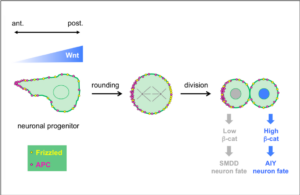
In a collaborative study published in Development, the teams of Vincent Bertrand and Pierre-François Lenne analyze the role played by Wnt ligands in the divisions that generate neurons during nervous system development.
Embryo development like a stadium wave
In a recent study appeared on the international journal Nature, Thomas Lecuit and his colleagues at the Institut de Biologie du Développement de Marseille describe how tissue shape changes are self-organized.
